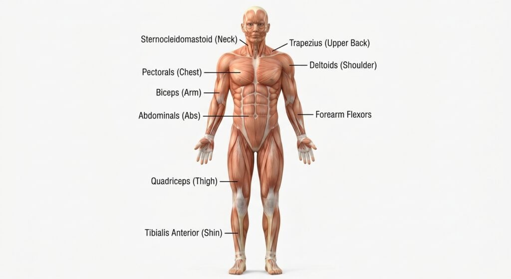
Muscles move your body by pulling on bones. A skeletal muscle gets a signal from a nerve, shortens, and tugs on a tendon, which swings a bone around a joint. Because a muscle can only pull, muscles work in opposing pairs: one bends a joint, and its partner straightens it again.
That’s the short answer to how do human muscles move the body, but the clever part happens where you can’t see it. Inside every muscle cell, tiny protein heads grab a neighboring filament, drag it about 10 nanometers, let go and grab again. Countless small tugs like that, spread across thousands of sarcomeres, add up to a wave, a jump or a pencil stroke.
By the end of this guide you’ll be able to explain the whole chain in an exam answer, from the thought “lift my arm” to the arm actually moving. You’ll also know the vocabulary teachers expect: tendon, origin, insertion, agonist, sarcomere, actin, myosin and ATP.
We’ll start with what a muscle does to a bone, then look inside a muscle, follow the seven steps of a contraction, see where the energy comes from, and finish with a worked example, common myths and quick FAQs.
Quick answer
Human muscles move the body by contracting and pulling on bones through tendons. A nerve signal makes each muscle fiber release calcium, which lets myosin heads pull actin filaments inward so the muscle shortens. Bones act as levers and joints act as pivots. Muscles only pull, so opposing pairs, like the biceps and triceps, move joints back and forth.
Key takeaways
- The body has about 600 muscles, and skeletal muscle is the type that moves your bones.
- A muscle can only pull, so every movement has a partner muscle that pulls the other way.
- Contraction runs on a chain: nerve signal, acetylcholine, calcium, then myosin pulling actin.
- ATP powers both the pull and the release; a muscle stores only a few seconds’ worth.
- Bones are levers, joints are the pivot points and muscles supply the effort.
What Does a Muscle Actually Do to Move a Bone?
A muscle moves a bone by shortening and pulling on it. It never pushes. Each skeletal muscle is anchored at two ends, and when it contracts, those two ends are drawn closer together, so one of the bones swings toward the other.
The connection is made by a tendon, a tough cord of collagen. Inside a muscle, the connective tissue wrapped around the fibers weaves into the tendon, and the tendon fuses with the outer layer of the bone. That’s why a strong contraction doesn’t simply tear the muscle off: the pull is spread through one continuous web of collagen.
Each muscle has two named attachment points:
- Origin: the end fixed to the bone that stays still.
- Insertion: the end fixed to the bone that moves.
Your biceps is a good one to feel. It’s anchored up near your shoulder and attaches to your forearm just below the elbow. Put your left hand on your right biceps and bend your right elbow: you’ll feel the muscle bunch up as it pulls the forearm (the insertion) toward the shoulder (the origin).
The skeleton gives muscles something solid to pull against. If you want to know which bones are doing the swinging, our guide on how many bones are in the human body breaks down the long bones of the arms and legs.
Why Do Muscles Work in Pairs?
Muscles work in pairs because a muscle can pull a bone in only one direction. To bring the bone back, a second muscle on the opposite side of the joint has to pull the other way.
At the elbow, the biceps brachii bends (flexes) the forearm and the triceps brachii, on the back of the upper arm, straightens (extends) it. The knee works the same way with the hamstrings bending it and the quadriceps straightening it. Biologists give each muscle a job title depending on the movement:
| Role | What it does | Example: bending the elbow |
|---|---|---|
| Prime mover (agonist) | Does most of the work for the movement | Biceps brachii |
| Synergist | Helps the prime mover | Brachialis, a muscle lying under the biceps |
| Antagonist | Has the opposite action; relaxes or brakes the movement | Triceps brachii |
| Fixator | Holds the anchor bone steady so the prime mover pulls efficiently | Muscles that steady the shoulder blade |
These titles swap when the movement changes. Straighten your elbow and the triceps becomes the prime mover while the biceps becomes the antagonist.
Antagonists aren’t just switched off, either. They help hold a limb in position and slow down fast movements, which is why you can throw a ball hard without your arm flinging itself out of the socket.
How Do Bones and Joints Work Like Levers?
Your skeleton moves like a set of levers. A bone is the rigid bar, a joint is the pivot (fulcrum), a muscle supplies the effort, and whatever is being moved (a body part or an object you’re holding) is the load.
Physics books describe three classes of lever, and the body has an example of each:
| Lever class | What sits in the middle | Body example | Fulcrum | Effort | Load |
|---|---|---|---|---|---|
| First class | The fulcrum | Tipping your head back | Joint between skull and top of spine | Neck muscles such as the trapezius | Weight of the head |
| Second class | The load | Rising onto your toes | Ball of the foot (toe joints) | Calf muscles, including the gastrocnemius | Body weight |
| Third class | The effort | Bending the elbow | Elbow joint | Biceps brachii | Forearm and anything in your hand |
The third-class lever at your elbow explains something odd about the biceps. It attaches close to the joint, so it must pull hard to lift even a light object. The payoff is range and speed: a muscle shortening by a few centimeters near the elbow moves your hand a much longer distance. Your body trades raw force for fast, wide movements.
The lever classes and their body examples come from an open anatomy textbook built on OpenStax:
LibreTexts Human Anatomy: Interactions of Skeletal Muscles and Their Lever Systems
What Are the Three Types of Muscle?
The body has three types of muscle tissue: skeletal, smooth and cardiac. Only skeletal muscle moves your bones, and it’s the only type you control on purpose.
| Type | Where you find it | Under your control? | Striped (striated) look? | Example job |
|---|---|---|---|---|
| Skeletal | Attached to bones by tendons (arms, legs, face, trunk) | Yes, voluntary | Yes | Walking, smiling, writing |
| Smooth | Walls of the stomach, intestines, bladder and blood vessels | No, involuntary | No | Squeezing food along the gut |
| Cardiac | Walls of the heart only | No, involuntary | Yes | Pumping blood with each beat |
Even “automatic” jobs can rely on skeletal muscle. Your diaphragm, the dome under your lungs, is skeletal muscle that keeps working while you sleep; our guide on how the lungs breathe shows how it pulls air in. Cardiac muscle gets its own story in how the human heart works.
How many muscles is that in total? The Better Health Channel, run by the Victoria State Government in Australia, puts the figure at about 600. Counts vary slightly because anatomists split some muscle groups differently.
Better Health Channel: Muscles
What Is Inside a Skeletal Muscle?
A skeletal muscle is built like a rope made of smaller ropes. Bundles sit inside bundles until you reach the protein filaments that do the actual pulling.
| Level | What it is | Wrapping or detail | Size |
|---|---|---|---|
| Whole muscle | An organ, such as the biceps | Wrapped in the epimysium | Varies |
| Fascicle | A bundle of muscle fibers | Wrapped in the perimysium | Many fibers per bundle |
| Muscle fiber | One long muscle cell with many nuclei | Wrapped in the endomysium | Up to 30 cm (11.8 in) long in the sartorius of the thigh |
| Myofibril | A strand running the full length of the fiber | Packed side by side in the fiber | About 1.2 micrometers wide |
| Sarcomere | The repeating unit that shortens | Lined up end to end along each myofibril | About 2 micrometers long |
| Filaments | Actin (thin) and myosin (thick) | Overlap inside each sarcomere | Molecular scale |
The sarcomere is the part to remember. It’s the smallest piece of muscle that can contract, and a myofibril is just thousands of sarcomeres linked in a chain. When every sarcomere along the chain shortens a little, the whole fiber shortens a lot.
Each fiber has its own nerve contact. A branch of a motor neuron meets the fiber at a spot called the neuromuscular junction, which is where the signal to contract arrives.
The sizes above are from OpenStax’s open college textbook:
OpenStax Anatomy and Physiology 2e: 10.2 Skeletal Muscle
What Happens Inside a Muscle When You Decide to Move?
When you decide to move, a nerve signal releases calcium inside your muscle fibers, and that calcium lets myosin pull on actin until the muscle shortens. Here’s the chain in seven steps, the order most biology exams expect.
- The brain sends the order. Your motor cortex plans a voluntary movement and the cerebellum helps keep it smooth. Each side of the motor cortex controls muscles on the opposite side of the body. For more on these brain areas, see how the human brain works.
- A motor neuron delivers the message. The signal travels down the spinal cord and out along a motor neuron. At the neuromuscular junction, the neuron releases a chemical messenger called acetylcholine.
- The muscle fiber fires. Acetylcholine lets sodium rush into the fiber, starting an electrical wave (an action potential). The wave races along the fiber’s surface and down tiny tunnels called T-tubules into its center.
- Calcium floods out. The electrical wave triggers the sarcoplasmic reticulum, the fiber’s internal calcium store, to release calcium ions.
- The binding sites open. Calcium attaches to a protein called troponin. Troponin then shifts a second protein, tropomyosin, which had been covering the grip points on the actin filaments.
- Myosin pulls. The myosin heads latch onto the exposed actin and swivel in a “power stroke,” dragging the actin about 10 nanometers toward the middle of the sarcomere. A fresh ATP molecule makes each head let go, and splitting that ATP re-cocks the head for the next grab. The cycle repeats as long as calcium and ATP are present.
- The muscle relaxes. When the nerve stops firing, pumps powered by ATP move calcium back into storage. Tropomyosin slides back over the grip points and the fiber goes slack.
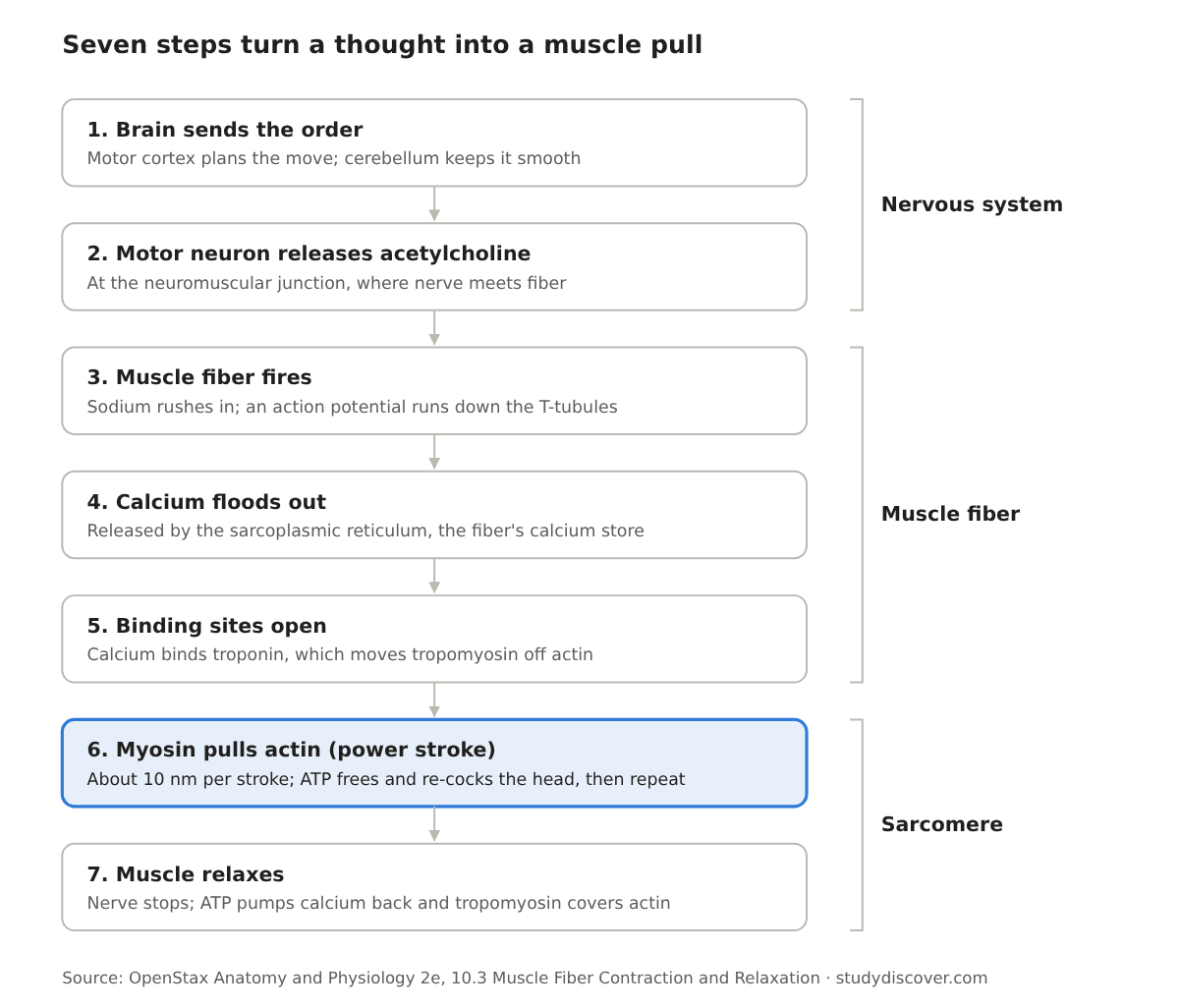
Picture of the seven steps of muscle contraction, from the brain’s signal to the muscle relaxing. Steps 1 and 2 happen in the nervous system, steps 3 to 5 in the muscle fiber, and steps 6 and 7 in the sarcomere.
Notice what doesn’t happen: the filaments themselves never get shorter. They slide past each other, so the overlap grows and the ends of each sarcomere (the Z lines) move closer together. Biologists call this the sliding filament model.
Every step above is described in OpenStax’s chapter on contraction:
OpenStax Anatomy and Physiology 2e: 10.3 Muscle Fiber Contraction and Relaxation
How Does the Brain Control How Hard a Muscle Pulls?
The brain controls force by choosing how many motor units to switch on. A motor unit is one motor neuron plus every muscle fiber it connects to, and all the fibers in one unit contract together.
Motor units come in very different sizes. The muscles that move your eyeballs have tiny units, with each neuron controlling only a handful of fibers, which allows precise little adjustments. Thigh and back muscles have large units, where one neuron can trigger thousands of fibers at once for raw power.
For a light task, such as picking up a sheet of paper, your nervous system activates a few small units. As the load grows, it adds more and larger ones. This step-by-step adding of units is called recruitment, and it’s why the same biceps can lift a feather or a full water bottle without you thinking about it.
Even at rest, a few units stay on in rotation. That low-level tension, called muscle tone, keeps muscles firm and helps hold your joints and posture steady.
Where Do Muscles Get the Energy to Move?
Muscles run on ATP (adenosine triphosphate), and they need it twice in every cycle: once to let myosin release actin and once to re-cock the myosin head. ATP also powers the pumps that pull calcium back during relaxation.
The catch is that a muscle stores only enough ATP for a few seconds of work. After that it has to rebuild ATP as fast as it spends it, using three systems that overlap:
| Energy source | Needs oxygen? | How long it can power activity | ATP made | Example activity (illustrative) |
|---|---|---|---|---|
| Stored ATP | No | A few seconds | Already made | The first moment of any movement |
| Creatine phosphate | No | About 15 seconds | Rebuilds ATP directly from ADP | A jump, a lift, a short sprint |
| Anaerobic glycolysis | No | About 1 minute | 2 ATP per glucose (net) | A hard 200 to 400 m run |
| Aerobic respiration | Yes | As long as fuel and oxygen last | About 36 ATP per glucose | Walking, steady jogging, resting muscle |
Aerobic respiration is slower but far more efficient. According to OpenStax, it supplies about 95 percent of the ATP used by resting or moderately active muscle. The no-oxygen route makes lactic acid as a by-product, which may add to the burning, tired feeling in a hard effort, though scientists still don’t know every cause of muscle fatigue.
What Are the Three Types of Muscle Contraction?
A muscle can produce force while getting shorter, while getting longer, or while staying the same length. All three are real contractions, and you use each one many times a day.
| Contraction type | What the muscle length does | Everyday example (biceps) |
|---|---|---|
| Concentric | Shortens while moving the load | Lifting a water bottle toward your shoulder |
| Eccentric | Lengthens while still under tension | Lowering the bottle back down slowly |
| Isometric | Stays the same; the joint angle doesn’t change | Holding the bottle still with your elbow bent at 90 degrees |
Eccentric work is the one students forget. Walking downstairs, sitting into a chair and setting down a heavy bag are all controlled by muscles lengthening as they resist gravity.
Worked Example: What Your Muscles Do When You Kick a Ball
Here’s an example scenario that puts every idea above into one movement: a right-footed kick at a soccer ball.
Step 1: The brain plans the kick
Your eyes track the ball and the left motor cortex (which runs the right side of your body) sends the command down your spinal cord. The cerebellum fine-tunes timing and balance.
Step 2: The wind-up
The hamstrings on the back of your right thigh contract concentrically and bend the knee, swinging your lower leg back. Their antagonists, the quadriceps on the front of the thigh, relax and stretch.
Step 3: The strike
The roles flip. The quadriceps become the prime movers and contract concentrically to snap the knee straight. Because the kick needs force, your nervous system recruits the large motor units of the thigh along with the small ones. Inside every fiber, calcium floods out and myosin heads cycle as fast as ATP allows.
Step 4: The follow-through
Once the foot meets the ball, the hamstrings work eccentrically: they lengthen under tension to brake the swinging leg so your knee isn’t over-straightened.
Step 5: Balance and fuel
Muscles in your left leg and hip contract isometrically to keep you upright on one foot. A kick is over in a moment, so it runs mostly on stored ATP and creatine phosphate rather than oxygen.
Try a slow-motion version while sitting down: put one hand on the front of your thigh and the other on the back, then swing your lower leg. You’ll feel the two muscle groups take turns.
Common Mix-Ups About Muscles
| Myth | Reality |
|---|---|
| Muscles can push bones as well as pull them. | Muscles only pull. Opposing pairs, such as the biceps and triceps, reverse a movement. |
| Actin and myosin filaments shrink when a muscle contracts. | The filaments keep their length and slide past each other; only the sarcomere gets shorter. |
| Muscles need ATP only to contract. | ATP is also needed for myosin to let go and for calcium to be pumped back. Without ATP, muscles lock, which is why bodies stiffen after death. |
| Lactic acid is the single cause of muscle fatigue. | It may contribute, but fatigue also involves falling ATP and ion imbalances, and its exact causes are not fully known. |
| You control all of your muscles. | Smooth muscle (gut, blood vessels) and cardiac muscle (heart) work without conscious control. |
Sources for the filament, ATP and fatigue rows:
OpenStax Anatomy and Physiology 2e: 10.3 Muscle Fiber Contraction and Relaxation
Source for the voluntary and involuntary row:
Better Health Channel: Muscles
Frequently Asked Questions
How many muscles are in the human body?
There are about 600 muscles in the human body, according to the Better Health Channel. You’ll see slightly different totals in different books because some muscles can be counted as one muscle or as several parts. Almost all of them are skeletal muscles; smooth and cardiac muscle form sheets in organ walls and the heart rather than separately named muscles.
Do muscles push or pull bones?
Muscles only pull. When a muscle contracts, it shortens and draws its two attachment points closer together. To move a bone back, a different muscle on the opposite side of the joint has to contract. That’s why muscles are arranged in pairs, such as the biceps and triceps at the elbow or the hamstrings and quadriceps at the knee.
What is the difference between skeletal, smooth and cardiac muscle?
Skeletal muscle is attached to bones, looks striped under a microscope and moves when you choose. Smooth muscle lines hollow organs like the stomach and blood vessels, has no stripes and works automatically. Cardiac muscle exists only in the heart; it’s striped like skeletal muscle but contracts on its own rhythm without your control.
Is a muscle an organ or just tissue?
Both words apply, at different levels. Muscle tissue is the type of tissue made of contracting cells. A named skeletal muscle, such as the biceps, counts as an organ because it combines muscle tissue with connective tissue wrappings, tendons, blood vessels and nerves that work together for one job.
What makes muscles get tired?
Muscles tire when they can’t keep up with the demands placed on them. Possible causes include ATP running low, lactic acid building up and making the inside of the cell more acidic, and shifts in sodium and potassium that disturb calcium release. Scientists still haven’t pinned down every cause.
Why do muscles stiffen after death?
Muscles stiffen after death (rigor mortis) because the body stops making ATP. Myosin heads need a fresh ATP molecule to release actin, so without it they stay locked onto the actin filaments. The muscles remain rigid until the proteins themselves begin to break down.
The Short Version
- Muscles move the body by contracting and pulling on bones through tendons; they never push.
- Opposing pairs, like the biceps and triceps, reverse each movement, and roles switch as the movement changes.
- A contraction runs from brain to motor neuron to acetylcholine to calcium to myosin pulling actin.
- ATP powers the pull, the release and relaxation; stored ATP lasts only seconds, so creatine phosphate, glycolysis and aerobic respiration refill it.
- Bones act as levers and joints as pivots, which trades force for speed and range.
Ready to check what stuck? Try the science quiz, or turn the seven contraction steps into cards with the Flashcard Maker. To see how muscles team up with the other organ systems, read how the human body works, or browse more guides in Biology.
For a deeper, college-level walk through how nerves control muscle force, the free OpenStax textbook is a reliable next stop:
OpenStax Anatomy and Physiology 2e: 10.4 Nervous System Control of Muscle Tension
Disclaimer
This article is for general educational purposes only. It explains how healthy muscles work and is not medical advice about muscle pain, injury or disease. For homework and exams, follow your teacher’s or exam board’s syllabus, and for any health concern, talk to a doctor or qualified health professional.